|
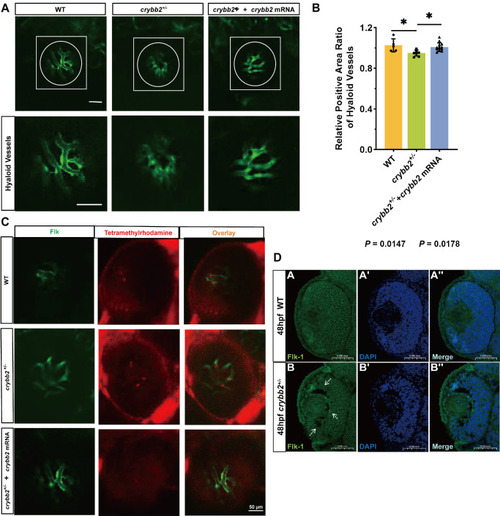
Crybb2 deficiency impairs hyaloid vessel development and permeability in zebrafish. (A) Confocal imaging of hyaloid vasculature in Tg(flk1:EGFP) zebrafish larvae at 5 dpf. White dashed lines outline vitreous vascular structures; boxed regions indicate magnified views. (B) Quantification of vascular density across experimental groups, analyzed using ImageJ. Each group included at least eight larvae (n ≥ 8). Statistical significance was determined using one-way ANOVA followed by Tukey's post-hoc multiple comparison test. Data are presented as mean ± SEM. (C) Assessment of tracer distribution by intravenous injection of TMR-MEP (red) at 5 dpf, imaged together with flk1:EGFP-positive vasculature (green). Scale bar: 50 µm. (D) Immunofluorescence staining for Flk-1 (VEGFR2/KDR, green) at 48 hpf. Nuclei were counterstained with DAPI (blue). Z-stack confocal images acquired at magnification ×60. Scale bar: 60 µm.
|