|
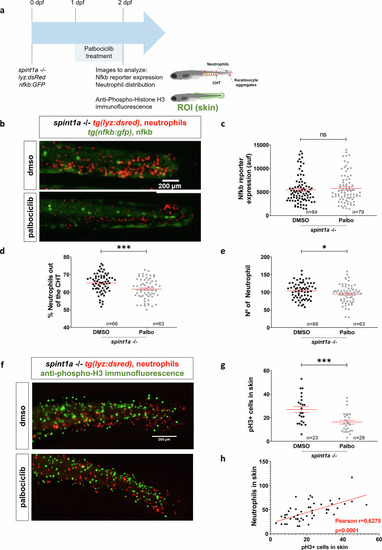
Keratinocyte proliferation is essential for facilitating neutrophil infiltration into inflamed skin. a Schematic of the experimental procedure used. Spint1a-deficient embryos were treated with Palbociclib or vehicle (DMSO) at 1 dpf by bath immersion. At 2 dpf, images were taken to analyze Nfkb reporter expression and neutrophil distribution. Anti-pH3 immunofluorescence was also performed. b Representative images of 2 dpf control and treated larvae, showing neutrophils and Nfkb reporter activity. c Quantification of Nfkb activity in skin in each group. Quantification of distribution (d) and number (e) of neutrophils in tail in every condition. e Quantification of neutrophil distribution in the tail in every condition. f Representative images of anti-pH3 immunofluorescence of 2 dpf control and treated larvae. g Number of pH3+ cells in skin in every condition. h Analysis of the correlation between the number of neutrophils and the number of pH3+ cells in skin. Each dot represents one individual, and the means and SEM for each group are also shown. P values were calculated by Student’s t test (c, d, f) and by Pearson Correlation test (g). ns, not significant; *P ≤ 0.05 ; ***P ≤ 0.001.
|