FIGURE

FIGURE 4

ID
ZDB-FIG-260105-66
Publication
Tseng et al., 2025 - Disruption of Swell1/VRAC function impairs initial hemodynamics and activates compensatory leukotriene signaling in zebrafish circulation development
Other Figures
All Figure Page
Back to All Figure Page
FIGURE 4

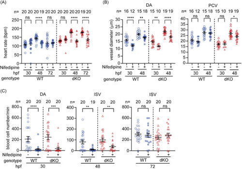
Inhibition of cardiac output by nifedipine further compromises hemodynamics in dKO embryos. (A) Nifedipine treatment significantly reduced the heart rate in WT embryos at 48 hpf and in dKO embryos at 48 and 72 hpf. (B) The vessel diameter of the dorsal aorta (DA) was significantly reduced in both WT and dKO embryos at 30 and 48 hpf following nifedipine treatment. The posterior cardinal vein (PCV) diameter was also significantly reduced in dKO embryos at 48 hpf. (C) Nifedipine treatment significantly reduced blood cell flow (hemodynamics) in both WT and dKO embryos at 30 and 48 hpf, but not at 72 hpf. The sample size (n) for each group is indicated above the scatter plot. The data were collected from two biological replicates. Nifedipine: vehicle control with 0.1% DMSO; +, 10 µM nifedipine. ns, no significance; *p < 0.05; **p < 0.01; ***p < 0.001; ****p < 0.0001.

Expression Data

Expression Detail
Antibody Labeling
Phenotype Data
Fish:
Condition:
Observed In:
Stage Range: Prim-15 to Protruding-mouth

Phenotype Detail
Acknowledgments
This image is the copyrighted work of the attributed author or publisher, and ZFIN has permission only to display this image to its users. Additional permissions should be obtained from the applicable author or publisher of the image. Full text @ Front Cell Dev Biol