FIGURE

FIGURE 2

ID
ZDB-FIG-260105-64
Publication
Tseng et al., 2025 - Disruption of Swell1/VRAC function impairs initial hemodynamics and activates compensatory leukotriene signaling in zebrafish circulation development
Other Figures
All Figure Page
Back to All Figure Page
FIGURE 2

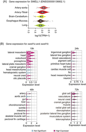
Tissue-specific expression of SWELL1 in humans and its orthologs swell1a and swell1b in zebrafish embryos. (A) Gene expression analysis of SWELL1 in human tissues based on data from the Genotype-Tissue Expression (GTEx) project. SWELL1 shows the highest expression in arterial tissues, including the aorta and tibial artery, relative to other tissues. (B) Spatial and temporal expression profiles of zebrafish swell1a and swell1b mined from scRNA-seq data. At 19 hpf, swell1a and swell1b exhibit high expression in cell types related to circulatory system development, including the lateral mesoderm, lateral plate mesoderm, and heart. By 24 hpf, expression remains elevated in blood vasculature and cardiac muscle cells. At 48 hpf, the highest expression is observed in arteries and the aortic arch. By 72 hpf, swell1a and swell1b expressions are enriched in vasculature and associated cell types. Cell types with significant expression levels are highlighted in pink, while non-significant expression is shown in blue.

Expression Data

Expression Detail
Antibody Labeling
Phenotype Data

Phenotype Detail
Acknowledgments
This image is the copyrighted work of the attributed author or publisher, and ZFIN has permission only to display this image to its users. Additional permissions should be obtained from the applicable author or publisher of the image. Full text @ Front Cell Dev Biol