IMAGE

FIGURE 4

ID
ZDB-IMAGE-260105-69
Source
Figures for Tseng et al., 2025
Image
Figure Caption

FIGURE 4

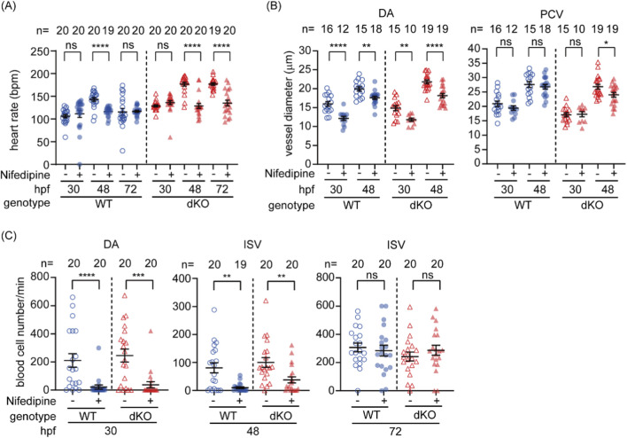
Inhibition of cardiac output by nifedipine further compromises hemodynamics in dKO embryos. (A) Nifedipine treatment significantly reduced the heart rate in WT embryos at 48 hpf and in dKO embryos at 48 and 72 hpf. (B) The vessel diameter of the dorsal aorta (DA) was significantly reduced in both WT and dKO embryos at 30 and 48 hpf following nifedipine treatment. The posterior cardinal vein (PCV) diameter was also significantly reduced in dKO embryos at 48 hpf. (C) Nifedipine treatment significantly reduced blood cell flow (hemodynamics) in both WT and dKO embryos at 30 and 48 hpf, but not at 72 hpf. The sample size (n) for each group is indicated above the scatter plot. The data were collected from two biological replicates. Nifedipine: vehicle control with 0.1% DMSO; +, 10 µM nifedipine. ns, no significance; *p < 0.05; **p < 0.01; ***p < 0.001; ****p < 0.0001.

Figure Data
Acknowledgments
This image is the copyrighted work of the attributed author or publisher, and ZFIN has permission only to display this image to its users. Additional permissions should be obtained from the applicable author or publisher of the image. Full text @ Front Cell Dev Biol