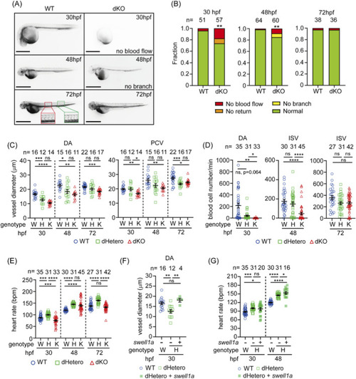
Hypovolemia and reduced hemodynamics in swell1-deficient embryos. (A) Microangiography revealed impaired circulation in dKO embryos. At 30 hpf, circulation was absent in 17.5% (10/57) of embryos, and in 8.8% (5/57), circulation did not return to the heart. Impaired circulation persisted in 8.3% (5/60) of dKO embryos at 48 hpf, with intersegmental vessels (ISVs) failing to develop in 6.67% (4/60) of embryos (no branching observed). By 72 hpf, impaired circulation was resolved in most dKO embryos. The red box indicates the region used to measure the average diameters of the dorsal aorta (DA) and posterior cardinal vein (PCV), and the green box marks the region used to calculate the number of blood cells flowing through the arterial ISVs. Scale bar: 100 μm. (B) Fraction bar diagram showing the circulatory system development in WT and dKO embryos. Sample sizes (n) are indicated above the bars. **p < 0.01. (C) The vessel diameter was measured from angiographic images. The DA was significantly smaller in dKO embryos at 30, 48, and 72 hpf and in dHetero embryos at 30 and 48 hpf, compared to WT embryos. The PCV diameter was also smaller in dKO embryos at 30, 48, and 72 hpf and in dHetero embryos at 72 hpf. (D) The number of blood cells flowing through the DA was significantly decreased in dHetero and dKO embryos at 30 hpf and through the intersegmental vessels (ISVs) in dKO embryos at 48 hpf. (E) Heart rate measurements show that dKO embryos have a slower heart rate than WT embryos at 30 hpf but a faster heart rate at 48 hpf. In dHetero embryos, the heart rate is consistently faster than in WT embryos at 30, 48, and 72 hpf. (F) Delayed circulation in dHetero embryos was rescued by swell1a mRNA injection. The vessel diameter of the DA in mRNA-injected dHetero embryos was comparable to WT embryos at 30 hpf. (G) The heart rate in swell1a mRNA-injected dHetero embryos was significantly faster than in WT embryos at 30 and 48 hpf. The sample size (n) for each group is indicated above the scatter plot. The data were collected from three biological replicates for (C–E) and from two biological replicates for (F, G). Genotypes: W (wild-type), H (dHetero), K (dKO). ns, no significance; *p < 0.05; **p < 0.01; ***p < 0.001, ****p < 0.0001.
|
