FIGURE

Figure 1

ID
ZDB-FIG-251122-2
Publication
Kuang et al., 2025 - BICD2 promotes ciliogenesis by facilitating CP110 removal from the mother centriole
Other Figures
All Figure Page
Back to All Figure Page
Figure 1

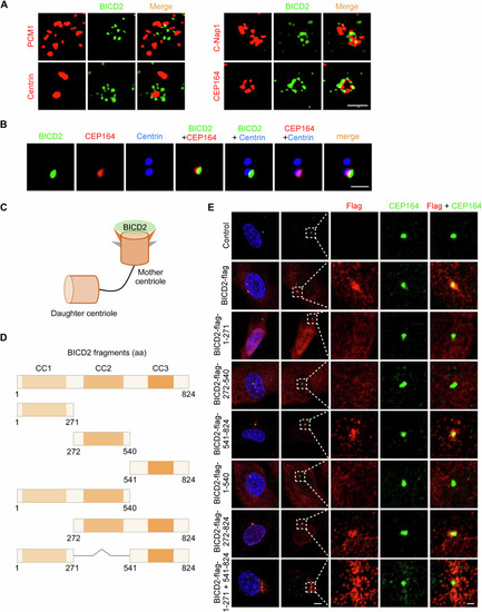
BICD2 is located at the mother centriole.

(A) 3D-SIM immunostaining images of RPE-1 cells stained with BICD2 antibody in conjunction with antibodies against PCM1, Centrin, C-Nap1, or CEP164. RPE-1 cells were cultured in normal serum medium. Scale bar, 1 μm. (B) Multi-color confocal images of RPE-1 cells stained with BICD2 antibody, along with Centrin and CEP164 antibodies. RPE-1 cells were cultured in normal serum medium. Scale bar, 1 μm. (C) Schematic illustration of BICD2 localization at the mother centriole. (D) Schematic diagrams of full-length BICD2 and BICD2 truncation mutants. (E) Immunostaining images of RPE-1 cells infected with lentiviruses carrying the indicated plasmids for 48 h, treated with puromycin for an additional 48 h, and stained with antibodies against Flag and CEP164. RPE-1 cells were cultured in normal serum medium. DNA was stained with DAPI. Scale bars, 5 μm (two columns on the left), 1 μm (three columns on the right). Source data are available online for this figure.

Expression Data

Expression Detail
Antibody Labeling
Phenotype Data

Phenotype Detail
Acknowledgments
This image is the copyrighted work of the attributed author or publisher, and ZFIN has permission only to display this image to its users. Additional permissions should be obtained from the applicable author or publisher of the image. Full text @ EMBO Rep.