|
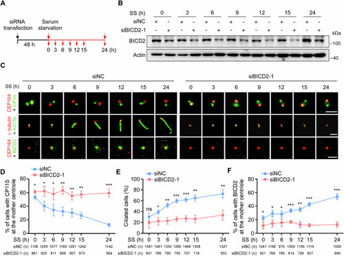
BICD2 is recruited to the mother centriole during cilia formation. (A–F) RPE-1 cells transfected with control or BICD2 siRNAs for 48 h were treated with serum starvation, and then subjected to Western blotting or immunofluorescence at the indicated time points. Schematic illustration of experimental strategy used for the cilia formation experiment (A). Western blot analysis of BICD2 protein at the indicated time points (B). Actin was used as a loading control. Immunofluorescence images of RPE-1 cells stained with antibodies against the indicated proteins (C). Scale bars, 2 μm. Quantification analyses of the percentage of cells with CP110 at the mother centriole (D), or ciliated cells (E), or cells with BICD2 at the mother centriole (F). CP110 at the mother centriole (D), P (SS0h, 3, 6, 9, 12, 15, 24 h) = 0.0165, 0.022, 0.0297, 0.0036, 0.0068, 0.0014, and 0.0002; Ciliated cells (E), P (SS0h, 3, 6, 9, 12, 15, 24 h) = 0.2029, 0.0224, 0.0025, 0.0005, 0.0008, 0.0022, and 0.0048; BICD2 at the mother centriole (F), P (SS0h, 3, 6, 9, 12, 15, 24 h) = 0.0389, 0.0238, 0.0373, 0.0008, 0.0017, 0.0001, 0.0002. n, the number of total cells calculated. Data were presented as mean ± SD from three independent biological repeats. Student’s t-test; ns not significant, *P < 0.05, **P < 0.01, ***P < 0.001. Source data are available online for this figure.
|