
Fig. 7
- ID
- ZDB-FIG-250916-28
- Publication
- Stefan et al., 2025 - Polypharmacology translates between species and phylogenetic distance: A functional, bioinformatic, and structural study on organic anion transporting polypeptides
- Other Figures
- All Figure Page
- Back to All Figure Page
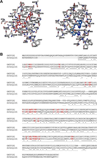
Structural comparison of the binding pockets of human OATP1B1 and drOatp1d1. A The protein backbones are shown in white (nitrogens: blue; oxygens: red; sulfurs: yellow), with key binding site residues labeled; left: BIL (red); right: PRA (blue). B Sequence alignment between human OATP1B1 (UniProt entry Q9Y6L6) and drOatp1d1 (UniProt entry A0A8M9PU70). The residues in the binding pocket highlighted in red, indicating conserved key residues involved in ligand interaction and transport function. (For interpretation of the references to color in this figure legend, the reader is referred to the web version of this article.) |