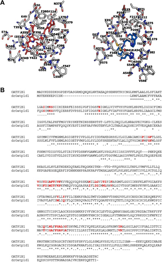
Fig. 7 Structural comparison of the binding pockets of human OATP1B1 and drOatp1d1. A The protein backbones are shown in white (nitrogens: blue; oxygens: red; sulfurs: yellow), with key binding site residues labeled; left: BIL (red); right: PRA (blue). B Sequence alignment between human OATP1B1 (UniProt entry Q9Y6L6) and drOatp1d1 (UniProt entry A0A8M9PU70). The residues in the binding pocket highlighted in red, indicating conserved key residues involved in ligand interaction and transport function. (For interpretation of the references to color in this figure legend, the reader is referred to the web version of this article.)
Image
Figure Caption
Acknowledgments
This image is the copyrighted work of the attributed author or publisher, and
ZFIN has permission only to display this image to its users.
Additional permissions should be obtained from the applicable author or publisher of the image.
Full text @ Biochem. Pharmacol.

