|
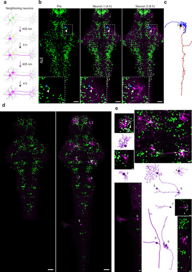
Pisces enables tracing the morphology of adjacent neurons and separated neurons across the brain. a Schematic illustrating the sequential activation of two neighboring neurons in the midbrain of 6-dpf larval zebrafish. b, c Fluorescent images showing the morphology of two tectal neurons activated sequentially. The first neuron (“1”) was designated by a white arrowhead, and the second (“2”) by a blue arrowhead (b). The dashed box highlights two sequentially activated neurons, shown in detail in the zoomed-in view below. Morphological traces of these two adjacent neurons are displayed in (c). Larvae were raised under ALE conditions. Scale bars: 50 μm in whole images, 10 μm in zoomed views. n = 6 fish. Representative z-axis maximum projection images (d), corresponding zoomed-in views, and morphological tracing (e) of nine neurons activated across the entire brain on 6-dpf larval zebrafish. Numbers indicate the activated neurons, which are shown in higher magnification in the zoomed-in panels on the right (n = 3 fish). Note that neuron 9 is not included in the pre-activation image. Larvae were raised in constant darkness. Scale bars: 50 μm in whole images, 10 μm in zoomed views. See also Supplementary Fig. 5, Supplementary Movies 7–9.
|