
Fig. 5
- ID
- ZDB-FIG-250714-26
- Publication
- Berger et al., 2024 - Sept10 and sept12 are expressed in specific proliferating cells in zebrafish brain
- Other Figures
- All Figure Page
- Back to All Figure Page
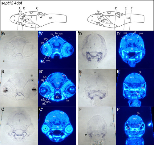
Transverse sections of 4 dpf zebrafish larvae showing detailed sept12 expression in the CNS. A – F: Epon cross sections of sept12 labeled 4 dpf zebrafish. Brain structures were visualized using Hoechst 33258 (A’ – F′). Levels and angles of sections are displayed in the schematic drawing. Note that the brain schemes display 5 dpf brains and serve only for rough orientation. Sept12 expression displayed in bright field images was prominent in cells at the border between tectum opticum (TeO) and cerebellar commissure (cec in C – D) as well as in a cell cluster at the medial ventricle (C–E). Sept12 was expressed in tissue outside the CNS (C–E). Scale bar: 100 μm. See list for abbreviations. Brain schemes were adapted from Mueller and Wullimann, 2016). |
| Gene: | |
|---|---|
| Fish: | |
| Anatomical Terms: | |
| Stage: | Day 4 |
Reprinted from Gene expression patterns : GEP, , Berger, C., Charlotte Kreß, J.K., Helmprobst, F., Sept10 and sept12 are expressed in specific proliferating cells in zebrafish brain, 119387, Copyright (2024) with permission from Elsevier. Full text @ Gene Expr. Patterns