
Fig. 2
- ID
- ZDB-FIG-250714-23
- Publication
- Berger et al., 2024 - Sept10 and sept12 are expressed in specific proliferating cells in zebrafish brain
- Other Figures
- All Figure Page
- Back to All Figure Page
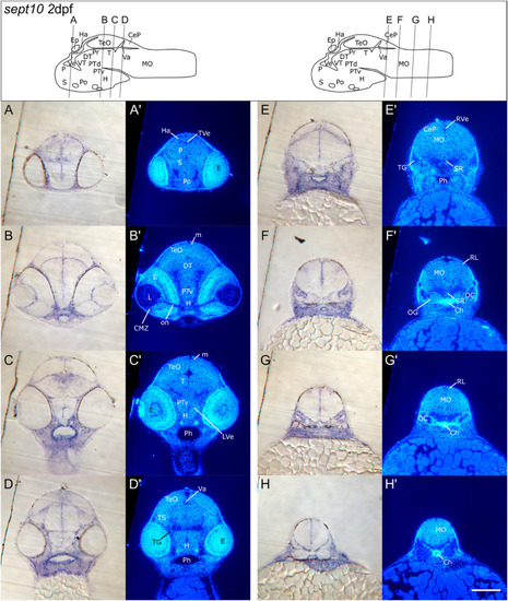
Transverse sections of 2 dpf zebrafish larvae showing detailed sept10 expression in the CNS. A–H: Transverse Epon sections of sept10 labeled 2 dpf zebrafish (A–H). Sections were counterstained with Hoechst 33258 to visualize brain structures (A’ - H′). Cutting levels and angles of the respective sections are illustrated in the brain schemes above. Bright field images show that sept10 expression was localized in ventricular zones as well as in several structures such as pallium (P), subpallium (S in A) and tegmentum (T in C). Sept10 expressing cells were also found around the lens (L), inside the optic nerve (on in B) and in regions of the cerebellar plate (CeP in E). Moreover, sept10 expression was observed in cells of the rhombic lip (RL) and medial in the medulla oblongata (MO) (F–H). Scale bar: 100 μm. See list for abbreviations. Brain schemes were adapted from Mueller and Wullimann, 2016). |
| Gene: | |
|---|---|
| Fish: | |
| Anatomical Terms: | |
| Stage: | Long-pec |
Reprinted from Gene expression patterns : GEP, , Berger, C., Charlotte Kreß, J.K., Helmprobst, F., Sept10 and sept12 are expressed in specific proliferating cells in zebrafish brain, 119387, Copyright (2024) with permission from Elsevier. Full text @ Gene Expr. Patterns