
Fig. 3
- ID
- ZDB-FIG-250714-24
- Publication
- Berger et al., 2024 - Sept10 and sept12 are expressed in specific proliferating cells in zebrafish brain
- Other Figures
- All Figure Page
- Back to All Figure Page
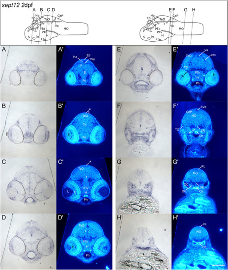
Transverse sections of 2 dpf zebrafish larvae showing detailed sept12 expression in the CNS. A–H: Transverse sections of sept12 labeled 2 dpf zebrafish (A–H). Sections were in addition stained with Hoechst 33258 for better visualization of distinct brain areas (A’ - H′). Level and angle of each section are displayed in the brain scheme above. Bright field images show sept12 expression in cells along the ventricle (A + B) and in several brain regions including habenula (Ha), pallium (P), subpallium (S in A), zones around the lens (L) and in the optic nerve (on in C). Sept12 expression was also visible at a dorsal region of the tectum opticum (TeO) (asterisk), tegmentum (T), hypothalamus (H) and posterior tuberculum ventral part (PTv in D). Furthermore, cells in the cerebellar comissure (cec), cerepellar plate (CeP), torus semicircularis (TS in E − F) and rhombic lip (RL) (G) showed sept12 expression. Prominent sept12 expression was also detected in the superior Raphe nucleus (SR) (F–G). Scale bar: 100 μm. See list for abbreviations. Brain schemes were adapted from Mueller and Wullimann, 2016). |
Reprinted from Gene expression patterns : GEP, , Berger, C., Charlotte Kreß, J.K., Helmprobst, F., Sept10 and sept12 are expressed in specific proliferating cells in zebrafish brain, 119387, Copyright (2024) with permission from Elsevier. Full text @ Gene Expr. Patterns