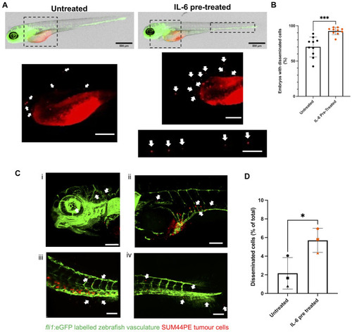
IL-6 promotes increased dissemination of SUM44PE cells in zebrafish embryos. (A) Representative images of 2dpi/4dpf Casper Tg(fli1:eGFP) Zebrafish embryos injected with untreated (left) or IL-6 pre-treated (right) SUM44PE cells, images on mesoscope at 3.2x, scale bar shows 500 μm with disseminated cells indicated by arrows. Dashed boxes represent zoomed in regions shown below, scale bars show 200 μm. (B) Quantification of the percentage of embryos with or without disseminated cells, n = 9 independent biological replicates. (C) Representative 3D projections of fixed 2dpi/4dpf embryos showing disseminated cells in the (i) head and heart, (ii) the SIV, (iii) CHT and (iv) the tail fin. Arrows indicate cells that have extravasated from the vasculature, z-stacks with 3 μm steps on Andor Dragonfly confocal microscope, scale bars show 100 μm. (D) Quantification of the percentage of all SUM44PE cells that are disseminated in the embryos (outside the primary site and extravasated from the vasculature). Quantified using Imaris 10.0.1. n = 3 biological replicates, with 3–4 embryos in each group per replicate, points represent the mean of each replicate. (C) and (E) two-way paired t-tests in GraphPad Prism, * p < 0.05, *** p < 0.001. fli1:eGFP labelled vasculature in green, DiI-dyed SUM44PE cells in red
|
