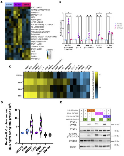
ILC CAF conditioned media drives STAT3 and MAPK pathway activation. (A) Heatmap of significantly changed proteins and phospho-proteins after 30 min–24 h of ED28 CAF conditioned media (CM) stimulation of SUM44PE cells compared to control cells. Protein expression was normalized to fast green stain for total protein and the heatmap shows the z-scores of expression. (B) Proteins with more than 2-fold and significant change in expression in the RPPA, determined by normalizing all values to mean of control. For (A) and (B), significance (* FDR < 0.05) was determined by two-way ANOVA, with multiple comparison correction by two-stage linear step-up procedure of Benjamini, Kreiger and Yeketel (BKY) in GraphPad Prism. (C) Heatmap of the top 20 most highly secreted proteins by primary ILC patient derived CAFs, n = 3 biological replicates for ED2334, ED26 and ED28, n = 2 for ED34. Heatmap shows log2 of signal intensity normalized to CAF cell pellet protein concentration, showing the mean across replicates. (D) Concentration of IL-6 in CM collected after 72 h from primary NST CAFs (blue), ILC CAFs (purple), and the ILC tumor cell lines SUM44PE and MM134 determined by ELISA, normalized to cell pellet protein concentration. (E) Western blot of SUM44PE cells stimulated for 30 min with recombinant human IL-6 (10 ng/mL) or CAF-CM from primary ILC CAFs (ED38 and ED2334) +/- anti-IL-6 blocking antibody (1 µg/mL)
|
