FIGURE

Fig. 4

ID
ZDB-FIG-250703-4
Publication
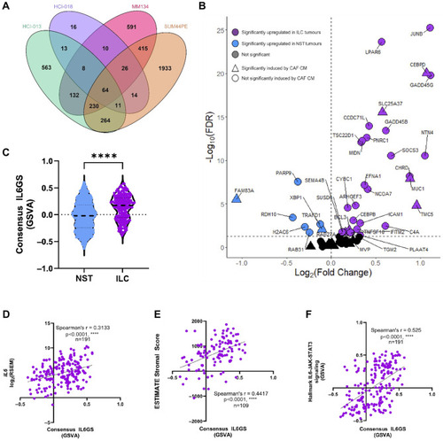
Bullock et al., 2025 - Cancer-associated fibroblast driven paracrine IL-6/STAT3 signaling promotes migration and dissemination in invasive lobular carcinoma
Other Figures
All Figure Page
Back to All Figure Page
Fig. 4

IL-6 drives consistent gene changes in multiple ILC models. (A) Venn diagram of differentially expressed genes in IL-6 treated HCI-013 (green), HCI-018 (purple) ER + ILC PDOs and MM134 (pink) and SUM44PE (orange) ILC cell lines. 64 genes were differentially expressed by IL-6 in all four models. (B) Volcano plot of differential expression of 61 genes in the consensus IL6GS between ER + ILC (n = 191) and ER + NST (n = 563) tumors in the TCGA Firehose dataset. Genes significantly differentially expressed (Wilcoxon U test, FDR < 0.05) and upregulated in ILC (purple) or NST (blue), triangles indicate genes also significantly upregulated by CAF CM in SUM44PE cells. (C) Consensus IL6GS score calculated by GSVA in ER + ILC and ER + NST tumors from TCGA, Mann-Whitney test **** p < 0.0001 in GraphPad Prism. Expression of the Consensus IL6GS score significantly and positively correlates with (D) IL6, (E) ESTIMATE stromal score (n = 109), and (F) Hallmark IL6-JAK-STAT3 signaling gene set (n = 191) in ER + ILC tumors, Spearman correlation in GraphPad Prism

Expression Data

Expression Detail
Antibody Labeling
Phenotype Data

Phenotype Detail
Acknowledgments
This image is the copyrighted work of the attributed author or publisher, and ZFIN has permission only to display this image to its users. Additional permissions should be obtained from the applicable author or publisher of the image. Full text @ Breast Cancer Res.