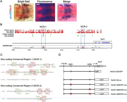
Noncoding conserved sequences found in the her3 upstream DNA. A. Comparison of the expression patterns in the neural plate between pou5f3 and her3. The expression of her3, stained red, was compared by two‐color WISH with that of pou5f3, stained in blue, at the bud stage. Dorsal views of flat‐mount preparations with anterior to the left. Broken lines mark the midbrain‐hindbrain boundary (mhb). Co‐expression of pou5f3 and her3 is marked with arrowheads. r1/2–4, rhombomeres 1/2–4. Scale bar, 100 μm. B. Comparison of the nucleotide sequences of the upstream 5.0‐kb DNA plus the coding regions between zebrafish her3 and spotted gar her3 using rVISTA. Prediction of the binding sequences for transcription factors (Oct, Sox, Nanog) were performed in parallel. Noncoding conserved regions (NCR‐1, NCR‐2) are marked with blue frames. Red vertical bars shown above indicate the zebrafish sequences to which transcription factors were predicted to bind, whereas green bars indicate the binding sites shared by the two species. C. Consensus transcription factor binding sites identified in the conserved sequences. Binding sites in the NCRs were searched by Match‐1.0 Public (vertebrates, cut‐off to minimize the sum of both error rates), and the sites for Oct (red) and Sox (green) are shown. ‘+’ and ‘–’ represent orientations of the binding sites. D. Structures of the constructs used in reporter assays. The genomic organization of her3 is shown at the top. The two NCRs are shown with blue ovals. Below are shown the structures of the EGFP and luciferase reporter constructs, in which the upstream 4.0‐kb DNA of her3 was ligated to the reporter genes. The EGFP reporter construct (Her3[−4.0]‐EGFP) was used for live imaging in embryos and cultured cells. The luciferase reporter Her3[−4.0]‐Luc and its deletion constructs, Her3[−4.0]dNCR1‐Luc, Her3[−4.0]dNCR2‐Luc, and Her3[−4.0]dNCRs‐Luc, were used in in vitro reporter assays.
|
