
Figure 7
- ID
- ZDB-FIG-250607-33
- Publication
- Chen et al., 2025 - KDM6A Deficiency Induces Myeloid Bias and Promotes CMML-Like Disease Through JAK/STAT3 Activation by Repressing SOCS3
- Other Figures
- All Figure Page
- Back to All Figure Page
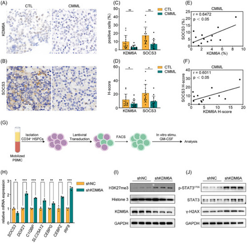
KDM6A/SOCS3/p‐STAT3 pathway is conserved in human HSPCs. A) Immunohistochemical staining of KDM6A on BM biopsies. B) Immunohistochemical staining of SOCS3 on BM biopsies. C) The percentage of KDM6A+ and SOCS3+cells in immunohistochemical staining. D) The H‐score of KDM6A+ and SOCS3+ cells immunohistochemical staining. E) Correlation analysis of the percentage of KDM6A+ (C, left) and SOCS3+ (C, right) cells in CMML specimens. F) Correlation analysis of the H‐score of KDM6A+ (D, left) and SOCS3+ (D, right) cells in CMML specimens. G) Schematic representation of human HSPC enrichment, lentiviral infection, and in vitro stimulation experiments conducted in this study. H) qPCR analysis of genes expression including |