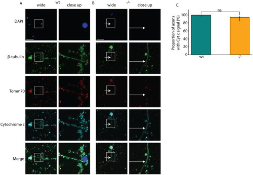
Mutation impacts the transport of mitochondria to the axons. The cultured brain neurons of wt and tomm70 mutant female fish were stained with DAPI (representing nucleus; blue), anti-β-Tubulin antibody (a neuronal marker; green), anti-Tomm70 antibody (red) and anti-Cytochrome c antibody (a conserved mitochondrial marker; cyan). The brightness of images was corrected using ImageJ. (A,B) Representative pictures of neuronal staining of wt (A) and mutant (B) fish with anti-β-Tubulin, anti-Tomm70 and anti-Cytochrome c antibodies. The white-line box in the wide-field column highlights the specific region magnified in the adjacent close-up column. White arrows in the mutants for β-Tubulin, Tomm70 and Cytochrome c staining indicate identical locations, underscoring the absence of Tomm70 and presence of Cytochrome c in these areas. Scale bars: 30 µm. (C) Quantification of the percentage of neuronal staining showing signal for Cytochrome c in the axons in wt and tomm70 mutants. Quantifications for Tomm70 signals are shown in Fig. 1G. N=4 (wt) and 3 (−/−); n=66 (wt) and 53 (−/−). Error bar represents 95% c.i. Statistical significance was tested using Fisher's permutation test. ns, non-significant.
|
