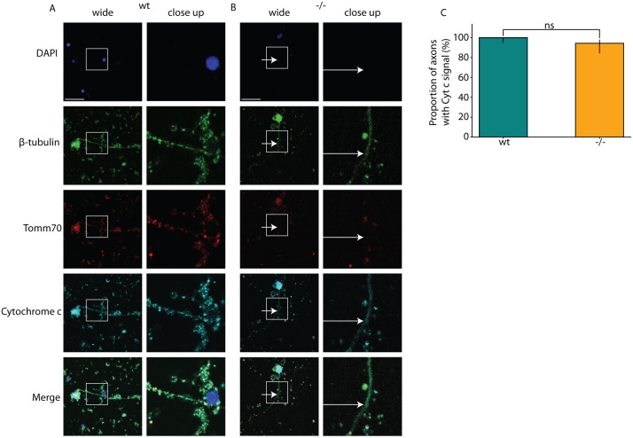
Mutation impacts the transport of mitochondria to the axons. The cultured brain neurons of wt and tomm70 mutant female fish were stained with DAPI (representing nucleus; blue), anti-β-Tubulin antibody (a neuronal marker; green), anti-Tomm70 antibody (red) and anti-Cytochrome c antibody (a conserved mitochondrial marker; cyan). The brightness of images was corrected using ImageJ. (A,B) Representative pictures of neuronal staining of wt (A) and mutant (B) fish with anti-β-Tubulin, anti-Tomm70 and anti-Cytochrome c antibodies. The white-line box in the wide-field column highlights the specific region magnified in the adjacent close-up column. White arrows in the mutants for β-Tubulin, Tomm70 and Cytochrome c staining indicate identical locations, underscoring the absence of Tomm70 and presence of Cytochrome c in these areas. Scale bars: 30 µm. (C) Quantification of the percentage of neuronal staining showing signal for Cytochrome c in the axons in wt and tomm70 mutants. Quantifications for Tomm70 signals are shown in Fig. 1G. N=4 (wt) and 3 (−/−); n=66 (wt) and 53 (−/−). Error bar represents 95% c.i. Statistical significance was tested using Fisher's permutation test. ns, non-significant.
This image is the copyrighted work of the attributed author or publisher, and
ZFIN has permission only to display this image to its users.
Additional permissions should be obtained from the applicable author or publisher of the image.
Full text @ Dis. Model. Mech.
Your Input Welcome
Thank you for submitting comments. Your input has been emailed to ZFIN curators who may contact you if
additional information is required.
Oops. Something went wrong. Please try again later.