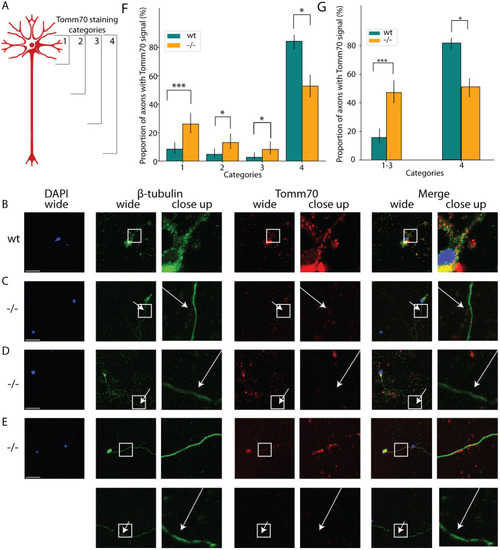
Mutation leads to the absence of Tomm70 from axons. The cultured brain neurons of wild-type (wt) and tomm70 mutant female fish were stained with DAPI (representing nucleus; blue), anti-β-Tubulin antibody (a neuronal marker) (green) and anti-Tomm70 antibody (red). The brightness of images was corrected using ImageJ. (A) The diagram represents four different categories of staining observed during imaging and quantification of the wt and tomm70 mutant brain cultured neurons stained with anti-Tomm70 antibody. Categories 1, 2, 3 and 4 depict the location of the signal for Tomm70 in the neurons. (B-E) Representative images of each category of Tomm70 staining in neurons. Scale bars: 30 µm. (B) wt fish showing signal for Tomm70 both in the soma and axon (category 4). (C) tomm70 fish showing signal for Tomm70 only in the soma (category 1). (D) tomm70 fish showing signal for Tomm70 in the soma and in the initial part of the axon (category 2). (E) tomm70 fish showing signal for Tomm70 in the soma and half way along the axon (category 3). The white-line box in the wide-field column highlights the specific region magnified in the adjacent close-up column. White arrows in the mutants for both β-Tubulin and Tomm70 staining indicate identical locations, underscoring the absence of Tomm70 in these areas. (F) Quantification representing the percentage of neuronal staining in four different categories for wt and mutants. More than 80% of the neuronal staining in wt was classified as category 4. Conversely, in the mutants, there was a significantly higher percentage of neuronal staining in categories 1, 2 and 3, and a significantly lower percentage of neuronal staining in category 4. (G) Quantification of the percentage of neuronal staining in categories 1-3 together, and for category 4, in wt and mutant fish. There is a significant increase in the percentage of neuronal staining in categories 1-3 in mutants compared to wt. Number of fish (N)=9 (wt) and 7 (−/−); total number of neurons counted (n)=191 (wt) and 146 (−/−). Error bar represents 95% c.i. Statistical significance was tested using Fisher's permutation test. *P<0.05 and ***P<0.001. ‘−/−’ in this and other figures refers to Danio rerio Tomm70Ile525Thr mutants, which possess a missense mutation, not a null mutation.
|
