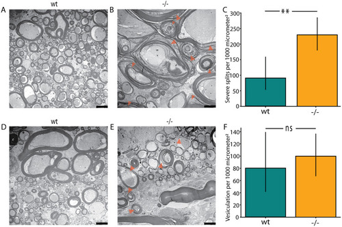
Myelin pathology in the large calibre axons of the spinal cord of tomm70 mutants. (A) Representative electron microscopy picture showing large calibre axons in the cranial part of the spinal cord with intact myelin in wt fish. (B) Orange arrowheads mark points of severe splitting of myelin sheath in the large calibre axons of the cranial part of the spinal cord in homozygous mutants. (C) Quantification of severe split events of myelin per 1000 µm2 area in wt and tomm70 mutant fish. There is a significant increase in the number of severe splitting cases in the cranial part of the spinal cord in mutants compared to wt. (D) Another electron micrograph showing intact myelin in the large calibre axons of the cranial part of the spinal cord in wt fish. (E) Orange arrowheads mark points of vesiculation of the myelin sheath surrounding the large calibre axons of the cranial part of the spinal cord in tomm70 mutant fish. (F) Quantification of vesiculation events of the myelin sheath per 1000 µm2 area in wt and mutant fish. Although there is a slight increase in the number of cases of vesiculation in mutants in the large calibre axons of the cranial part of spinal cord, it is not changed significantly compared to that in wt. N=2 (wt) and 5 (−/−). Error bar represents 95% c.i. Statistical significance was tested using Fisher's permutation test. **P<0.01; ns, non-significant. Scale bars: 2500 nm.
|
