
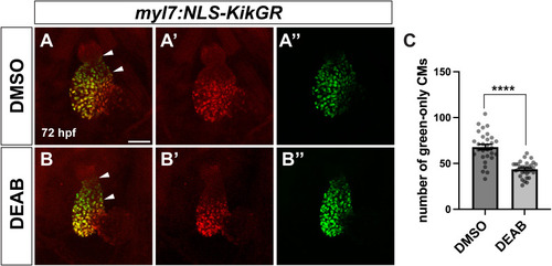
Fig. 4
SHF addition to the arterial pole of the ventricle is reduced in late RA-deficient embryos. (A, B) Representative images of temporal differentiation assay using the myl7:NLS-KikGR transgene in control DMSO (A) and DEAB-treated (B) embryos. White arrowheads indicate region of green-only later-differentiating cardiomyocytes of the SHF at the arterial pole. (A′, B′) Red channel only. (A″, B″) Green channel only. Maximum intensity projections of a Z-stack are shown for each image. Scale bar is 50 μM. (C) Quantification of green-only cardiomyocytes at 72 hpf in control (n = 30) and DEAB-treated (n = 32) embryos. Error bars represent SEM. ∗∗∗∗ indicates p < 0.0001. (For interpretation of the references to colour in this figure legend, the reader is referred to the Web version of this article.) |
Reprinted from Developmental Biology, , Griffin, A.H.C., Small, A.M., Johnson, R.D., Medina, A.M., Kollar, K.T., Nazir, R.A., McGuire, A.M., Schumacher, J.A., Retinoic acid promotes second heart field addition and regulates ventral aorta patterning in zebrafish, , Copyright (2025) with permission from Elsevier. Full text @ Dev. Biol.