
Fig. 2
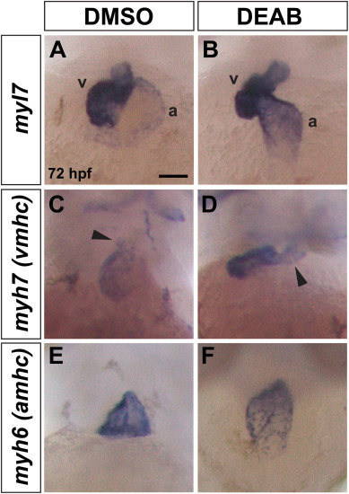
Heart morphology is abnormal in late RA-deficient embryos. (A, B) myl7 expression is similar in the ventricle (v) and atrium (a) of control (n = 35/35 have normal morphology as shown) and DEAB-treated embryos (n = 27/37 have elongated, linear morphology as shown, 10/37 have more horizontal chamber placement as seen in controls). (C, D) myh7 expression is present in the ventricle of DEAB-treated embryos (n = 31), but the ventricle is more compact with an elongated OFT region compared to the controls (n = 23). For control embryos, 21/23 have short OFT as shown, 2/23 have elongated OFT. For DEAB-treated embryos, 10/31 have short OFT, 21/31 have elongated OFT as shown. (E, F) myh6 expression is present in the atrium of DEAB-treated embryos but the chamber morphology is abnormal compared to controls. For control embryos, 48/48 have normal morphology as shown. For DEAB-treated embryos, 32/39 have variable abnormal elongated morphology. All embryos are 72 hpf. Scale bar is 50 μM. |
Reprinted from Developmental Biology, , Griffin, A.H.C., Small, A.M., Johnson, R.D., Medina, A.M., Kollar, K.T., Nazir, R.A., McGuire, A.M., Schumacher, J.A., Retinoic acid promotes second heart field addition and regulates ventral aorta patterning in zebrafish, , Copyright (2025) with permission from Elsevier. Full text @ Dev. Biol.