
FIGURE
Fig. 1
Fig. 1
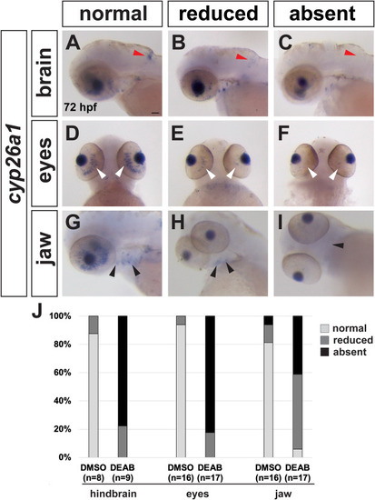
Cyp26a1 expression is reduced in embryos treated with DEAB from 26 hpf to 72 hpf. cyp26a1 expression in the hindbrain (A-C, red arrowheads), eyes (D-F, white arrowheads) and jaw (G-I, black arrowheads) is strongly reduced or absent in DEAB-treated embryos. J shows quantification of expression in each domain. All embryos are 72 hpf. Scale bar is 50 μM. (For interpretation of the references to colour in this figure legend, the reader is referred to the Web version of this article.) |
Expression Data
Expression Detail
Antibody Labeling
Phenotype Data
Phenotype Detail
Acknowledgments
This image is the copyrighted work of the attributed author or publisher, and
ZFIN has permission only to display this image to its users.
Additional permissions should be obtained from the applicable author or publisher of the image.
Reprinted from Developmental Biology, , Griffin, A.H.C., Small, A.M., Johnson, R.D., Medina, A.M., Kollar, K.T., Nazir, R.A., McGuire, A.M., Schumacher, J.A., Retinoic acid promotes second heart field addition and regulates ventral aorta patterning in zebrafish, , Copyright (2025) with permission from Elsevier. Full text @ Dev. Biol.