
FIGURE
Fig. 3
Fig. 3
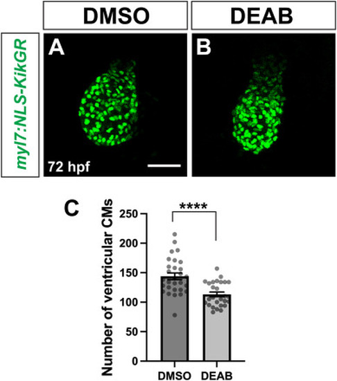
Ventricular cardiomyocyte number is reduced in late RA-deficient embryos. (A, B) Representative images of my7:NLS-KikGR expression in the ventricle of control DMSO (A) and DEAB-treated (B) embryos at 72 hpf. Note that expression is absent in the atrium, preventing analysis of that chamber. Maximum intensity projections of a Z-stack are shown for each image. Scale bar is 50 μM. (C) Number of ventricular cardiomyocytes at 72 hpf in control (n = 29) and DEAB-treated (n = 27) embryos. Error bars represent SEM. ∗∗∗∗ indicates p < 0.0001. |
Expression Data
Expression Detail
Antibody Labeling
Phenotype Data
Phenotype Detail
Acknowledgments
This image is the copyrighted work of the attributed author or publisher, and
ZFIN has permission only to display this image to its users.
Additional permissions should be obtained from the applicable author or publisher of the image.
Reprinted from Developmental Biology, , Griffin, A.H.C., Small, A.M., Johnson, R.D., Medina, A.M., Kollar, K.T., Nazir, R.A., McGuire, A.M., Schumacher, J.A., Retinoic acid promotes second heart field addition and regulates ventral aorta patterning in zebrafish, , Copyright (2025) with permission from Elsevier. Full text @ Dev. Biol.