
Fig. 2
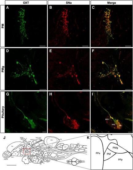
Immunocytochemical localization of Oxt-ir and Scg2a/SNa-ir in female zebrafish. Microscopy imaging of Oxt-ir (green) and Scg2a/SNa-ir (red) preoptic magnocellular (PM; A–C) and gigantocellular (PMg; D–F) regions, and pituitary (G–I). Merged images show colocalization (yellow-orange) of both immunoreactivities in these paraffin sections. RPD, rostral pars distalis; PPD, proximal pars distalis; NIL, neurointermediate lobe. A–F) 20× magnification; G–I) 10× magnification. The position of the PM and PMg relative to the zebrafish forebrain (J), and the PPa and PPa (anterior and posterior parts of parvocellular preoptic nucleus) are shown for reference. Scale bars in A–I = 50 µm. Scale bar in J = 500 µm |