
Fig. 5
- ID
- ZDB-FIG-250403-20
- Publication
- El-Hage et al., 2025 - Schwann cells have a limited window of time in which to initiate myelination signaling during early migration in vivo
- Other Figures
- All Figure Page
- Back to All Figure Page
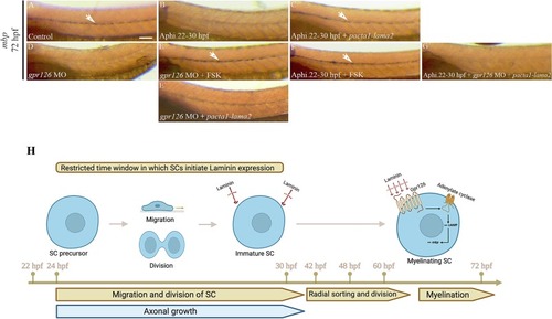
Laminin signals through Gpr126/cAMP to restore myelination in aphidicolin treated embryos. Lateral views of mbp expression at 3 dpf revealed by in situ hybridization along the PLLn in control showing a robust expression (A, n = 32/32), aphidicolin treated embryos showing a sharp decrease in mbp expression (B, n = 30/34), aphidicolin treated embryo and injected with pacta-lama2 (C, n = 37/38) showing a normal mbp expression in clones of SCs. gpr126 morphants show a significant reduction in mbp expression (D, n = 30/32) while those treated with FSK show a normal expression of mbp (E, n = 27/30) similar to embryos treated with aphidicolin and FSK (F, n = 31/33). Gpr126 morphants injected with pacta1-lama2 show a significant reduction in mbp expression (E′, n = 22/26). Embryos treated with aphidicolin and injected with pacta1-lama2 fail to express mbp in the absence of Gpr126 (G, n = 30/34). Arrows indicate SCs expressing mbp along the PLLn. Scale bar = 200 μm. (H) Model of SC development, data strongly suggest a model in which SCs initiate Laminin expression during migration. A gradual increase in the expression level and polymerization of Laminin drives radial sorting and myelination through Gpr126/cAMP signaling at later stages. |