|
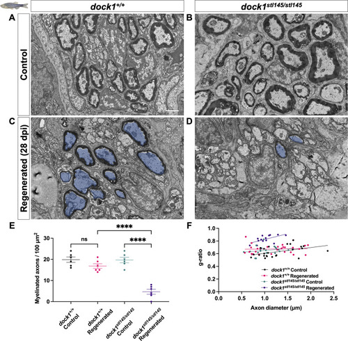
Remyelination following nerve injury is significantly reduced in dock1 MUTs. (A and B) TEM micrographs of control ZMBs from 4-mo-old WT dock1+/+ and MUT dock1stl145/stl145 zebrafish. (C and D) TEM micrographs of WT dock1+/+ and MUT dock1stl145/stl145 zebrafish showing regeneration and remyelination after transection, with remyelinated axons pseudocolored in blue. (E) Quantification of the number of myelinated axons in the ZMBs per 100 µm2, n = 6 (WT dock1+/+ control), 6 (WT dock1+/+ regenerated), 6 (dock1stl145/stl145 MUT control), and 6 (dock1stl145/stl145 MUT regenerated). (F) Quantification of the g-ratio as it relates to the axon caliber of the remyelinated axons in the regenerated ZMBs, 28 days after transection, n = 6 (WT dock1+/+ control), 6 (WT dock1+/+ regenerated), 6 (dock1stl145/stl145 MUT control), and 6 (dock1stl145/stl145 MUT regenerated). (A–D) Scale bar = 1 µm. (E) Two-way ANOVA with Tukey’s multiple comparisons test. ****P < 0.0001; ns, not significant.
|