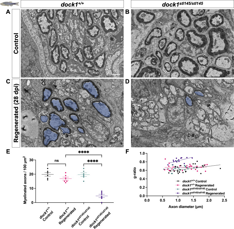
Remyelination following nerve injury is significantly reduced in dock1 MUTs. (A and B) TEM micrographs of control ZMBs from 4-mo-old WT dock1+/+ and MUT dock1stl145/stl145 zebrafish. (C and D) TEM micrographs of WT dock1+/+ and MUT dock1stl145/stl145 zebrafish showing regeneration and remyelination after transection, with remyelinated axons pseudocolored in blue. (E) Quantification of the number of myelinated axons in the ZMBs per 100 µm2, n = 6 (WT dock1+/+ control), 6 (WT dock1+/+ regenerated), 6 (dock1stl145/stl145 MUT control), and 6 (dock1stl145/stl145 MUT regenerated). (F) Quantification of the g-ratio as it relates to the axon caliber of the remyelinated axons in the regenerated ZMBs, 28 days after transection, n = 6 (WT dock1+/+ control), 6 (WT dock1+/+ regenerated), 6 (dock1stl145/stl145 MUT control), and 6 (dock1stl145/stl145 MUT regenerated). (A–D) Scale bar = 1 µm. (E) Two-way ANOVA with Tukey’s multiple comparisons test. ****P < 0.0001; ns, not significant.
This image is the copyrighted work of the attributed author or publisher, and
ZFIN has permission only to display this image to its users.
Additional permissions should be obtained from the applicable author or publisher of the image.
Full text @ J. Cell Biol.
Your Input Welcome
Thank you for submitting comments. Your input has been emailed to ZFIN curators who may contact you if
additional information is required.
Oops. Something went wrong. Please try again later.