
FIGURE
Fig. 1
- ID
- ZDB-FIG-250311-53
- Publication
- Mulzer et al., 2025 - Dynamic changes of extracellular vesicles during zebrafish organogenesis
- Other Figures
- All Figure Page
- Back to All Figure Page
Fig. 1
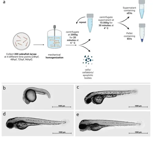
Protocol of EV isolation and zebrafish larvae development: a: Isolation protocol for whole zfl sEVs and lEVs by differential centrifugation. b-e: Zfl development; Imaging was performed at the 4 observation time points in 2% low-melting agarose (peqLab, Erlangen, Germany) using an Axio Observer microscope system (Zeiss, Jena, Germany), magnification 2,5x. b: 24 hpf, c: 48 hpf, d: 72 hpf, e: 96 hpf |
Expression Data
Expression Detail
Antibody Labeling
Phenotype Data
Phenotype Detail
Acknowledgments
This image is the copyrighted work of the attributed author or publisher, and
ZFIN has permission only to display this image to its users.
Additional permissions should be obtained from the applicable author or publisher of the image.
Full text @ Cell Commun. Signal.