
Fig. 4
- ID
- ZDB-FIG-250311-56
- Publication
- Mulzer et al., 2025 - Dynamic changes of extracellular vesicles during zebrafish organogenesis
- Other Figures
- All Figure Page
- Back to All Figure Page
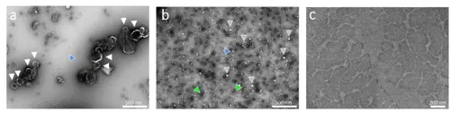
Morphology characterization of whole zfl sEVs and lEVs by TEM: The micrographs were collected on a JEOL 1400 Plus TEM (JEOL Germany, Munich, Germany) operating at 120 kV with a nominal magnification of 30.000x. a: 10.000 g homogenized whole zfl pellet at 30k magnification: cup shaped lEVs (white arrowhead), in the background some triangular shaped protein (blue arrowhead). The white bar in the lower right quadrant of the image corresponds to 500 nm. b: 10.000 g homogenized whole zfl supernatant at 30k magnification: sEVs of around ~ 70–100 nm (some depicted with grey arrowheads), the background is covered with triangular shaped protein (blue arrowhead) and some proteins aggregates appear (two marked with green arrowheads). The white bar in the lower right quadrant of the image corresponds to 500 nm. c: Zfl medium at 72 hpf at 30k magnification: no EVs, background with staining artefact. The white bar in the lower right quadrant of the image corresponds to 200 nm |