FIGURE

Fig. 3

ID
ZDB-FIG-250225-79
Publication
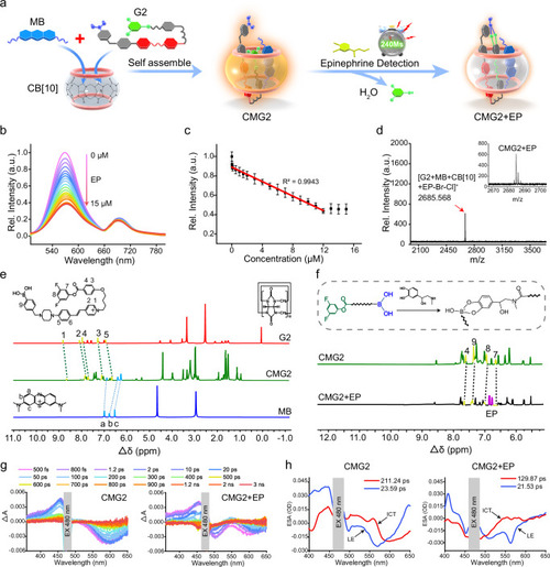
Zhao et al., 2025 - Molecularly engineered supramolecular fluorescent chemodosimeter for measuring epinephrine dynamics
Other Figures
All Figure Page
Back to All Figure Page
Fig. 3

Fluorescence titration of CMG2 toward EP and mechanism evaluation.

a The working principle of the developed and optimized CMG2 for specific and rapid determination of EP. b Fluorescence spectra of 10.0 μm CMG2 with the addition of EP at different concentrations (0– 15 μm) in cell lysis buffer (10 mm, pH = 7.4) containing 0.05% DMSO excited at 480 nm. c Relative fluorescence intensity of the CMG2 versus EP concentration (0–15 μm). Data are presented as mean ± S.D. Error bars: S.D., n  =  5 independent experiments. d Maldi-TOF mass spectrometry of CMG2 + EP. e1HNMR spectra of G2, MB and CMG2 ([G2] = [MB] = [CB10] = [CMG2] = 5 mm). f1HNMR spectra of CMG2 and CMG2 assembled with EP ([CMG2] = [EP] = 5 mm, in DMSO-d6: D2O = 1: 1 at 298 K). g Transient absorption spectroscopy for CMG2 (left), and CMG2 + EP (right). h Principal spectral components from the global analysis of time-resolved absorption spectra from 480 nm excitation for CMG2 (left), and CMG2 + EP (right). The above-mentioned source data are provided as a Source Data file.

Expression Data

Expression Detail
Antibody Labeling
Phenotype Data

Phenotype Detail
Acknowledgments
This image is the copyrighted work of the attributed author or publisher, and ZFIN has permission only to display this image to its users. Additional permissions should be obtained from the applicable author or publisher of the image. Full text @ Nat. Commun.