FIGURE

Fig 2

ID
ZDB-FIG-250207-62
Publication
Widziolek et al., 2025 - Gingipains protect Porphyromonas gingivalis from macrophage-mediated phagocytic clearance
Other Figures
All Figure Page
Back to All Figure Page
Fig 2

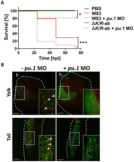
Myeloid cell-depleted zebrafish larvae are more susceptible to Porphyromonas gingivalis (Pg) infection.

(A) Kaplan-Meyer survival plot of pu.1 MO-treated or PBS-injected control zebrafish larvae upon systemic infection with the wild-type Pg W83 or ΔK/R-ab. Survival plots were analysed by log-rank test.*p≤0.05,***p≤0.001; hpi- hours post infection. (B) Representative images of the yolk and tail region of pu.1 MO-treated and untreated zebrafish larvae 2 hpi with fluorescein-SE (green) and pHrodo (red) dual-stained ΔK/R-ab. The untreated larvae display orange dots (white arrows) representing phagocytosed (phagosome acidified) ΔK/R-ab. Phagocytosed ΔK/R-ab were not detected in pu.1 MO due to removal of myeloid cells. White dashed lines indicate yolk or tail areas. Boxes show zoomed regions of the images. h-heart. Scale bar = 100 μm.

Expression Data

Expression Detail
Antibody Labeling
Phenotype Data

Phenotype Detail
Acknowledgments
This image is the copyrighted work of the attributed author or publisher, and ZFIN has permission only to display this image to its users. Additional permissions should be obtained from the applicable author or publisher of the image. Full text @ PLoS Pathog.