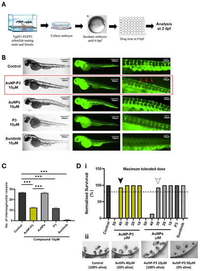
Pyrazinib-functionalised gold nanoparticles (AuNP-P3) inhibited developmental angiogenesis in Tg(fli1:EGFP) zebrafish. (A) Intersegmental vessel assay schematic; 6 h post-fertilization (hpf) Tg(fli1) embryos were treated with compounds and fixed and analyzed at 2 days post-fertilization (dpf). (B) Representative fluorescent images illustrating GFP-positive intersegmental vessels at low and high magnification and brightfield images of whole larvae at 2 dpf in Tg(fli1) zebrafish. Then, 6 hpf embryos were treated with the control (0.1% DMSO) C, pyrazinib-functionalised gold nanoparticles (AuNP-P3) 10 μM, AuNPs (gold nanoparticles) 10 μM, pyrazinib (P3) 10 μM, and sunitinib 10 μM as indicated on the graph. Red arrows indicate the altered vessel formation observed with AuNP-P3 treatment. (C) The number of intersegmental vessels was quantified at 2 dpf. One-way ANOVA with Tukey’s multiple comparisons test. Data are expressed as mean ± SEM; *** p < 0.001 (n = 15). (D) (i) Maximum tolerated dose (MTD) of the compounds in the Tg(fli1) embryos. Then, 6 hpf embryos were treated with 0.1% DMSO control, AuNP-P3 10, 20, 30, 40, and 50 μM, AuNPs (gold nanoparticles) 10, 20, 30, 40, and 50 μM, pyrazinib (P3) 10 μM, and sunitinib 10 μM. The dotted line denotes the 80% survival rate used as a cut-off for the establishment of the MTD. The survival of zebrafish treated with AuNP-P3 exceeded 80% at concentrations ≤ 40 μM (black arrow), while at concentrations ≤ 30 μM (white arrow) for AuNPs (n = 15). (ii) Representative images of live and dead zebrafish larvae at 2 dpf.
|
