FIGURE SUMMARY
Title

Targeting Radiation Resistance in Oesophageal Adenocarcinoma with Pyrazinib-Functionalised Gold Nanoparticles

Authors
Marcone, S., Spadavecchia, J., Khan, M., Vella, G., O'Connell, F., Pendino, M., Menon, M., Donohoe, C., Narayanasamy, R., Reynolds, J.V., Maher, S.G., Lynam-Lennon, N., Kennedy, B., Prina-Mello, A., O'Sullivan, J.
Source
Full text @ Cancers

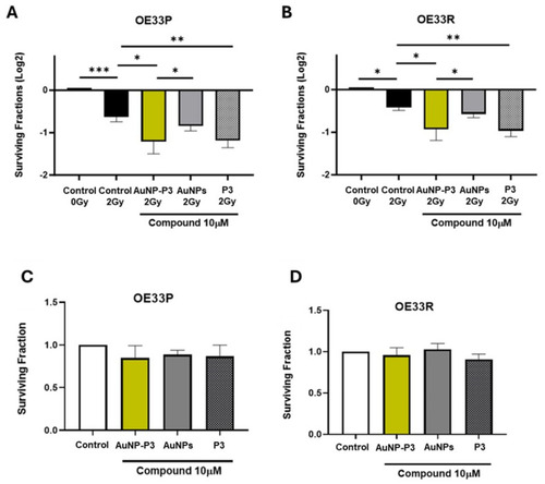
Pyrazinib-functionalised gold nanoparticles (AuNP-P3) enhanced radiosensitivity in an the OAC cell line model of radioresistance. The effect of pyrazinib-coupled gold nanoparticles (AuNP-P3), pyrazinib (P3), and gold nanoparticles (AuNPs) at 10 µM treatment on the radiosensitivity of OE33P (radiation sensitive) and OE33R (radioresistant) cells was assessed by clonogenic assays and compared to the control (0.1% DMSO + 0.1% water). Surviving fraction of OE33P (A) and OE33R (B) cells following treatment with compounds at 10 µM and 2 Gy X-ray radiation. Clonogenic assay was used to assess cell survival based on the ability of a single cell to grow into a colony after receiving treatment with the compound at 10 µM. Cell viability was assessed in mock-radiated OE33P (C) and OE33R cells (D). Data are expressed as mean ± SEM. Statistical analysis was carried out using a two-tailed paired t-test. * p < 0.05, ** p < 0.01, *** p < 0.001 (n = 4).

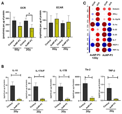
Pyrazinib-functionalised gold nanoparticles (AuNP-P3) treatment reduced oxidative phosphorylation and glycolysis in response to ionizing radiation in an OAC cell line model of radioresistance. OE33P (radiation sensitive cells) and OE33R (radiation resistant cells) were treated for 24 h with the compounds at 10 µM or with the control (0.1% DMSO + 0.1% water) and subsequently irradiated at 2 Gy X-ray radiation. After 24 h, cells were used to measure metabolic rates and the supernatants were stored for multiplex ELISA. Oxygen consumption rate (OCR) and extracellular acidification rate (ECAR) were measured in real-time using Seahorse Biosciences XFe24 analyser. Comparison of basal and radiation-induced OCR (A) and ECAR (B) levels between OE33P and OE33R cells. Relative percentage OCR (C,D) and ECAR (E,F) in OE33P cells following treatment with compounds at 10 μM and mock or 2 Gy radiation. Relative percentage OCR (G,H) and ECAR (I,J) in OE33R cells following treatment with compounds at 10 μM and mock or 2 Gy radiation. Data are expressed as mean ± SEM. Statistical analysis was carried out using an unpaired t-test to compare different cell lines and paired t-test to compare within the same cell line. * p < 0.05, ** p < 0.01 (Compound vs. Control); # p < 0.05 (AuNP-P3 vs. AuNPs) (n = 3).

Pyrazinib-functionalised gold nanoparticles (AuNP-P3) significantly altered the levels of secreted mediators in response to ionizing radiation in an OAC cell line model of radioresistance. The secreted levels of protein in OE33P (radiation sensitive) and OE33R (radiation resistant) cells were evaluated by 54-plex ELISA. Here, we report data only for the significantly modulated proteins. (A) AuNP-P3 10 µM significantly alters the level of 9 proteins (IL-2, IL-7, IL-12p40, IL12-p70, IL-13, IL-16, IL1β, MCP-1, and sICAM-1) in OE33P cells (radiosensitive) when compared to the control (0.1% DMSO). (B) AuNP-P3 10 µM significantly alters the level of 5 proteins (IL-2, IL-10, IL12-p70, IL-16, and IL1β) in OE33R cells (radioresistant) when compared to the control (0.1% DMSO + 0.1% water). Two-tailed paired t-test; * p < 0.05, ** p < 0.01, *** p < 0.001, # p < 0.05 (P3 vs. AuNP-P3) (n = 3). Data are expressed as mean ± SEM (n = 3). (C) Correlation plots showing significant correlations between the levels of secreted proteins and the metabolic rates in OE33P and OE33R cells treated with AuNP-P3 10 µM in the presence or absence of 2 Gy radiation. (Spearman correlation, blue indicates positive correlations and red indicates inverse negative correlations). The Holm–Bonferroni hoc correction was used to control for multiple comparison testing. * p < 0.05, ** p < 0.01, *** p < 0.001.

Pyrazinib-functionalised gold nanoparticles (AuNP-P3) decreased metabolic parameters and released mediators in OAC tumour explants in response to ionizing radiation. OAC explants were treated with pyrazinib-functionalised gold nanoparticles (AuNP-P3) at 10 µM, and after 18 h incubation, explants were exposed to 2 Gy X-ray radiation. After 6h incubation, OCR and ECAR were measured in real-time. Tissue-conditioned media were collected after Seahorse measurement and used for multiplex ELISA analysis. (A) AuNP-P3 significantly inhibits OCR in OAC explants exposed to 2 Gy radiation, when compared to the control (0.1% water). ANOVA with Šídák’s multiple comparisons test, * p  <  0.05. Data expressed as mean  ±  SEM (n = 3). (B) Altered released cytokines in AuNP-P3 treated OAC explants subjected to 2 Gy X-ray radiation were measured by multiplex ELISA. Two-tailed unpaired t-test; * p < 0.5. Data expressed as mean  ±  SEM (n = 3). (C) Correlation plots showing significant correlations between the levels of secreted proteins and the metabolic rates in huma OAC fresh explant treated with AuNP-P3 10µM in the presence or absence of 2 Gy radiation. Spearman correlation; blue indicates positive correlations and red indicates inverse negative correlations. The Holm–Bonferroni hoc correction was used to control for multiple comparison testing. * p < 0.05, ** p < 0.01.

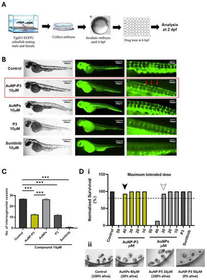
Pyrazinib-functionalised gold nanoparticles (AuNP-P3) inhibited developmental angiogenesis in Tg(fli1:EGFP) zebrafish. (A) Intersegmental vessel assay schematic; 6 h post-fertilization (hpf) Tg(fli1) embryos were treated with compounds and fixed and analyzed at 2 days post-fertilization (dpf). (B) Representative fluorescent images illustrating GFP-positive intersegmental vessels at low and high magnification and brightfield images of whole larvae at 2 dpf in Tg(fli1) zebrafish. Then, 6 hpf embryos were treated with the control (0.1% DMSO) C, pyrazinib-functionalised gold nanoparticles (AuNP-P3) 10 μM, AuNPs (gold nanoparticles) 10 μM, pyrazinib (P3) 10 μM, and sunitinib 10 μM as indicated on the graph. Red arrows indicate the altered vessel formation observed with AuNP-P3 treatment. (C) The number of intersegmental vessels was quantified at 2 dpf. One-way ANOVA with Tukey’s multiple comparisons test. Data are expressed as mean ± SEM; *** p < 0.001 (n = 15). (D) (i) Maximum tolerated dose (MTD) of the compounds in the Tg(fli1) embryos. Then, 6 hpf embryos were treated with 0.1% DMSO control, AuNP-P3 10, 20, 30, 40, and 50 μM, AuNPs (gold nanoparticles) 10, 20, 30, 40, and 50 μM, pyrazinib (P3) 10 μM, and sunitinib 10 μM. The dotted line denotes the 80% survival rate used as a cut-off for the establishment of the MTD. The survival of zebrafish treated with AuNP-P3 exceeded 80% at concentrations ≤ 40 μM (black arrow), while at concentrations ≤ 30 μM (white arrow) for AuNPs (n = 15). (ii) Representative images of live and dead zebrafish larvae at 2 dpf.

Acknowledgments
This image is the copyrighted work of the attributed author or publisher, and ZFIN has permission only to display this image to its users. Additional permissions should be obtained from the applicable author or publisher of the image. Full text @ Cancers