IMAGE

Figure 5

ID
ZDB-IMAGE-241219-86
Source
Figures for Marcone et al., 2024
Image
Figure Caption

Figure 5

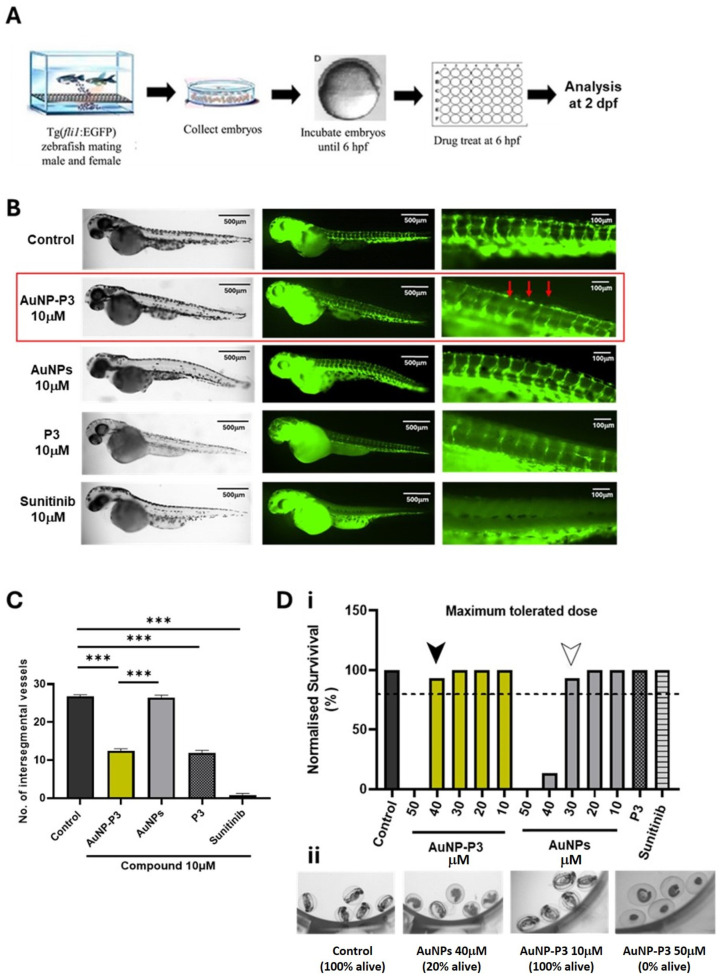
Pyrazinib-functionalised gold nanoparticles (AuNP-P3) inhibited developmental angiogenesis in Tg(fli1:EGFP) zebrafish. (A) Intersegmental vessel assay schematic; 6 h post-fertilization (hpf) Tg(fli1) embryos were treated with compounds and fixed and analyzed at 2 days post-fertilization (dpf). (B) Representative fluorescent images illustrating GFP-positive intersegmental vessels at low and high magnification and brightfield images of whole larvae at 2 dpf in Tg(fli1) zebrafish. Then, 6 hpf embryos were treated with the control (0.1% DMSO) C, pyrazinib-functionalised gold nanoparticles (AuNP-P3) 10 μM, AuNPs (gold nanoparticles) 10 μM, pyrazinib (P3) 10 μM, and sunitinib 10 μM as indicated on the graph. Red arrows indicate the altered vessel formation observed with AuNP-P3 treatment. (C) The number of intersegmental vessels was quantified at 2 dpf. One-way ANOVA with Tukey’s multiple comparisons test. Data are expressed as mean ± SEM; *** p < 0.001 (n = 15). (D) (i) Maximum tolerated dose (MTD) of the compounds in the Tg(fli1) embryos. Then, 6 hpf embryos were treated with 0.1% DMSO control, AuNP-P3 10, 20, 30, 40, and 50 μM, AuNPs (gold nanoparticles) 10, 20, 30, 40, and 50 μM, pyrazinib (P3) 10 μM, and sunitinib 10 μM. The dotted line denotes the 80% survival rate used as a cut-off for the establishment of the MTD. The survival of zebrafish treated with AuNP-P3 exceeded 80% at concentrations ≤ 40 μM (black arrow), while at concentrations ≤ 30 μM (white arrow) for AuNPs (n = 15). (ii) Representative images of live and dead zebrafish larvae at 2 dpf.

Acknowledgments
This image is the copyrighted work of the attributed author or publisher, and ZFIN has permission only to display this image to its users. Additional permissions should be obtained from the applicable author or publisher of the image. Full text @ Cancers