
Fig 1
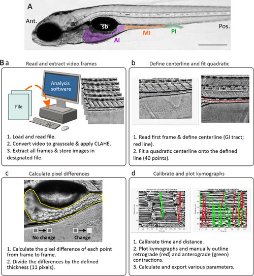
The kymography protocol. (A) Bright-field image of a lateral view of a zebrafish larva at 7 dpf showing the anterior intestine (AI, purple), mid-intestine (MI, orange) and posterior intestine (PI; green). Sb is swim bladder. Ant. And Pos. are anterior and posterior, respectively. Scale bar: 200 μm. (B) Schematic to demonstrate the four main steps (Ba-Bd) used to analyse the imaging data via kymography. In (Bd), the green and red lines are indications of contractions in the anterograde and retrograde direction, respectively. There were drawn using line tools available in MATLAB (R2018b, MathWorks, Natick, MA, USA). |