
Fig. 6
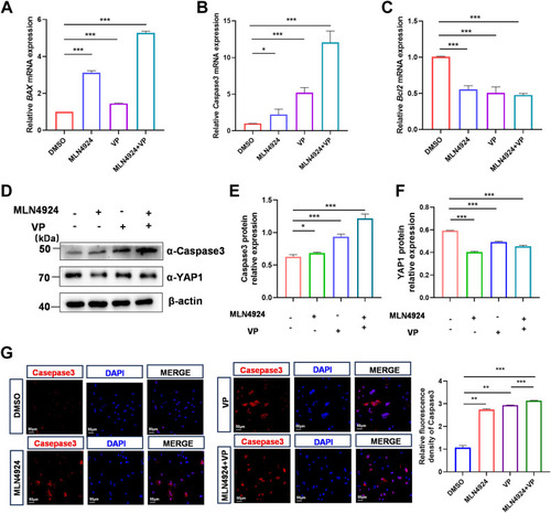
Effect of YAP1 suppression on MLN4924-induced apoptosis in rabbit GCs. A–C, qPCR analysis of proapoptotic marker genes Bax (A), Caspase3 (B), and antiapoptotic marker gene Bcl2 (C) mRNA levels in GCs treated with DMSO, MLN4924 (1 μM), VP (1 μM), or MLN4924 and VP together for 24 h. D–F, GCs were treated with DMSO, MLN4924 (1 μM), VP (5 μM), or MLN4924 and VP together for 24 h. Cell lysates were used for Western blot, and then YAP1 and Caspase3 protein levels were quantified and normalized to that of β-actin. A representative Western blot (D), the quantification of YAP1 (E) and Caspase3 (F) from three independent experiments. G, representative immunofluorescence staining for Caspase3 (red) after DMSO, MLN4924 (1 μM), VP (5 μM), or MLN4924 and VP together for 48 h. Quantification analysis of relative Caspase3 expression level (fluorescence intensity of Caspase3/DAPI) by Image J software. Data are presented as mean ± SD, ∗p < 0.05, ∗∗p < 0.01, and ∗∗∗p < 0.001. DAPI, 4′, 6-diamidino-2-phenylindole; DMSO, dimethyl sulfoxide; GC, granulosa cell; ns, not significant; qPCR, quantitative real-time PCR; VP, verteporfin; YAP1, yes-associated protein 1. |