
Fig. 3
- ID
- ZDB-FIG-240812-15
- Publication
- Guo et al., 2024 - Lysosomal membrane protein TMEM106B modulates hematopoietic stem and progenitor cell proliferation and differentiation by regulating LAMP2A stability
- Other Figures
- All Figure Page
- Back to All Figure Page
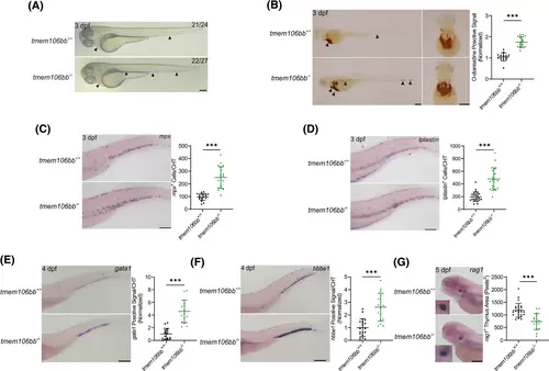
KO of tmem106bb impairs definitive hematopoiesis. (A) Appearance of embryos showed increased red blood cells in tmem106bb−/− embryos at 3 dpf (n = 24–27 embryos per group; scale bar: 200 μm). The black arrowheads indicate red blood cells. (B) O-dianisidine staining showed a significantly increased signal for red blood cells in tmem106bb−/− embryos at 3 dpf (n = 15–18 embryos per group; scale bar: 200 μm). The black arrowheads indicate red blood cells. (C) WISH for mpx showed that myeloid differentiation was significantly increased in tmem106bb−/− embryos at 3 dpf (n = 19–20 embryos per group; scale bar: 200 μm). (D) WISH for lplastin showed that myeloid differentiation was significantly increased in tmem106bb−/− embryos at 3 dpf (n = 17–26 embryos per group; scale bar: 200 μm). (E) WISH for gata1 showed that erythroid differentiation was significantly increased in tmem106bb−/− embryos at 4 dpf (n = 16-embryos per group; scale bar: 200 μm). (F) WISH for hbbe1 showed that erythroid differentiation was significantly increased in tmem106bb−/− embryos at 4 dpf (n = 16–20 embryos per group; scale bar: 200 μm). (G) WISH for rag1 showed that lymphoid differentiation was significantly reduced in tmem106bb−/− embryos at 5 dpf (n = 16–26 embryos per group; scale bar: 200 μm). ***p < .001; Data were shown as mean ± SD; Statistical analysis was performed using a Shapiro–Wilk test for samples with normal distribution and unpaired student's t tests with two tails. |