
Fig. 6
- ID
- ZDB-FIG-240620-14
- Publication
- Singh et al., 2024 - Forward genetics combined with unsupervised classifications identified zebrafish mutants affecting biliary system formation
- Other Figures
- All Figure Page
- Back to All Figure Page
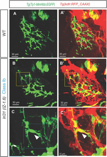
Biliary epithelial cells segregate from the intrahepatic biliary network in lri31 (IZ-1.6) mutant larvae at 5 dpf. (A–C) Projected confocal images of wild-type (A) and lri31 (IZ-1.6) mutant (B and C) larvae visualized for Tg(Tp1-MmHbb:EGFP)um14 expressions at 5 dpf. Tg(kdrl:RFP_CAAX)y171 expressions are merged in (A′-C′). Yellow-outlined area in B is magnified and shown in C. Blue-outlined area in C is magnified and merged with the DAPI and GFP co-localization signal in the left bottom corner. Ventral views, anterior to the top. In lri31 (IZ-1.6) mutant larvae, single biliary epithelial cells segregate from the intrahepatic biliary network and make physical contact with the intrahepatic vascular network (White arrowhead in C). DAPI and GFP co-localization assays revealed that six BECs are segregated from the main biliary tree in this larva. |
Reprinted from Developmental Biology, 512, Singh, D.J., Tuscano, K.M., Ortega, A.L., Dimri, M., Tae, K., Lee, W., Muslim, M.A., Rivera Paz, I.M., Liu, J.L., Pierce, L.X., McClendon, A., Gibson, I., Livesay, J., Sakaguchi, T.F., Forward genetics combined with unsupervised classifications identified zebrafish mutants affecting biliary system formation, 44-56, Copyright (2024) with permission from Elsevier. Full text @ Dev. Biol.