|
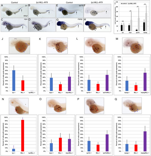
MLL expression colocalized with mpx, spi1b, and mpeg expression on the yolk of lyz:MLL-AF9 injected embryos at 48 and 72 hpf. Expression of lyz, mpx, spi1b, and mpeg in control (A,C,E,G) and lyz:MLL-AF9 injected (B,D,F,H) embryos at 48 hpf. Black arrows indicate expression in the CHT, arrowheads indicate expression on the yolk (A-H). The number of cells on the yolk expressing lyz, mpx, spi1b, and mpeg was assessed at 48 hpf (I, N = 31–62). MLL colocalization with lyz (0%), mpx (41.9% +/- 14.0%), spi1b (47.1% +/-14.1%), and mpeg (53.4% +/- 13.6%) was determined at 48 hpf (J-M, N = 37–52) and with lyz (0%), mpx (36.6% +/- 15.4%), spi1b (49.8% +/- 12.0%), and mpeg (58.8% +/- 16.0%) at 72hpf (N-Q, N = 26–34). Blue, red, and purple bars indicate the percentage of cells stained on the yolk that were myeloid single positive (blue arrow), MLL single positive (red arrow), or myeloid marker/MLL double positive (purple arrow), respectively (J-Q). Statistical analysis was conducted using student’s t-test. * p<0.05, ** p<0.01, NS = not significant. Anterior to the left.
|