
Fig. 1
- ID
- ZDB-FIG-240529-50
- Publication
- Sun et al., 2024 - ptx3a+ fibroblast/epicardial cells provide a transient macrophage niche to promote heart regeneration
- Other Figures
- All Figure Page
- Back to All Figure Page
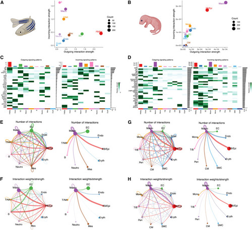
Tight communication between Fb/Epi cells and macrophages during zebrafish and neonatal mice heart regeneration (A) Scatterplot showing the overall incoming and outgoing communication strength based on receptor and ligand expression in the regenerating zebrafish heart. Circle size represents the total number of interactions associated with each cell cluster. (B) Scatterplot showing the overall incoming and outgoing communication strength based on receptor and ligand expression in the regenerating neonatal mouse heart. Circle size represents the total number of interactions associated with each cell cluster. (C) Heatmap showing the summary of the signaling pathways that contribute to outgoing or incoming communication in the regenerating zebrafish heart. The color bar represents the relative signaling strength of a signaling pathway across cell types. The bars indicate the sum of the signaling strength of each cell type. (D) Heatmap showing the summary of the signaling pathways that contribute to outgoing or incoming communication in the regenerating neonatal mouse heart. The color bar represents the relative signaling strength of a signaling pathway across cell types. The bars indicate the sum of the signaling strength of each cell type. (E and F) Circle plot showing differential cell-cell communication networks in the regenerating zebrafish heart. The width of the edges represents the relative number of interactions (E) or interaction strength (F). Arrow indicates target cell. (G and H) Circle plot showing differential cell-cell communication networks in the regenerating neonatal mouse heart. The width of the edges represents the relative number of interactions (G) or interaction strength (H). Arrow indicates target cell. |