
Fig. 1
- ID
- ZDB-FIG-240528-74
- Publication
- Ruparelia et al., 2024 - Atrogin-1 promotes muscle homeostasis by regulating levels of endoplasmic reticulum chaperone BiP
- Other Figures
- All Figure Page
- Back to All Figure Page
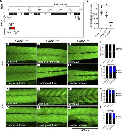
Atrogin-1 deficiency results in contraction-dependent muscle fiber detachment. Schematic of wild-type atrogin-1 (atrogin-1+/+) and mutant atrogin-1 (atrogin-1–/–) protein structure and mRNA sequence, with the mutant predicted to incorporate a premature stop in exon 1. The mutant was generated using CRISPR/Cas9 genome editing, which resulted in a 34 bp insertion (red). Numbers in the protein box are amino acids, and numbers in the mRNA box are base pairs. (B) qRT-PCR analysis showing significant reduction in atrogin-1 levels in atrogin-1–/– mutants compared with atrogin-1+/+ wild-type larvae. Error bars represent mean ± SEM for 3 replicate experiments, with each experiment comprising a pooled sample of at least 5 fish. *P < 0.05 determined using a 1-way ANOVA with Tukey’s multiple correction post hoc test. Muscle fibers span the entire length of the somite in the 3 dpf atrogin-1+/+ (C), atrogin-1 heterozygous (atrogin-1+/–) (D), and atrogin-1–/– mutant (E) larvae, as seen by F-Actin labeling. (F) Quantification of the muscle phenotype, with atrogin-1+/+, atrogin-1+/–, and atrogin-1–/– displaying indistinguishable muscle structure, as determined using a χ2 test. Incubation of 3 dpf atrogin-1+/– (H) and atrogin-1–/– (I) in methyl cellulose results in muscle fiber detachment, which is not evident in atrogin-1+/+ larvae (G). (J) Percentage of affected atrogin-1+/+, atrogin-1+/–, and atrogin-1–/– larvae, with the latter 2 genotypes having a significant increase in the proportion of fish displaying the muscle fiber detachment, as determined using a χ2 test. At 6 dpf, atrogin-1+/– (L) and atrogin-1–/– (M) display sporadic muscle fiber detachment but not in atrogin-1+/+ larvae (K). (N) Percentage of affected atrogin-1+/+, atrogin-1+/–, and atrogin-1–/– larvae, with the latter 2 genotypes having a significant increase in the proportion of fish displaying the muscle fiber detachment, as determined using a χ2 test. Methyl cellulose incubation of 6 dpf atrogin-1+/– (P) and atrogin-1–/– (Q) results in muscle fiber detachment, which is not evident in atrogin-1+/+ larvae (O). (R) Percentage of affected atrogin-1+/+, atrogin-1+/–, and atrogin-1–/– larvae, with the latter 2 genotypes having a significant increase in the proportion of fish displaying the muscle fiber detachment, as determined using a χ2 test. *P < 0.05, **P < 0.01, ***P < 0.001, ****P < 0.0001. All experiments were performed in triplicate, with the total number of fish examined in each replicate documented in Supplemental Table 2. |