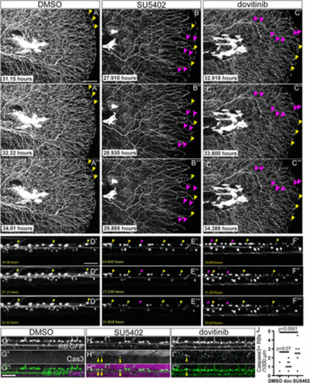
Fig. 8
|
Fgf inhibition and dovitinib treatment induce Wallerian-like axon degeneration and RB neuron apoptosis. A–C, Stills from time-lapse movies of larval RB:GFP caudal tails treated with DMSO (A), SU5402 (B), or dovitinib (C). Yellow arrowheads = maintained axon terminals, magenta arrowheads = degenerating axons. D–F, Stills from time lapse movies of larval RB:GFP dorsal neurons demonstrating RB apoptosis. Yellow arrowheads = surviving RB neurons, magenta arrowheads = RB neurons undergoing apoptosis. G–I, Lateral view of RB:GFP dorsal neurons immunostained for cleaved Caspase-3 to mark apoptosis (yellow arrowheads). Compared to DMSO-treated larvae (G), SU5402 (H), and dovitinib (I). Scale bars = 50 μm. I, treatment displayed Cas3+ RB neurons. J, Quantification of RB neuron apoptosis detected by Cas3+ immunostaining. DMSO = 0 ± 0.2 RB neurons, dovitinib = 1.2 ± 0.2, SU5402 = 2.6 ± 0, analyzed by one-way ANOVA with post hoc Tukey's test (F = 07.81, p = 0.0013). Scale bars = 50 μm. |

