Fig. 6
|
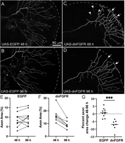
Cell autonomous loss of Fgf signaling in RB neurons induces loss of axon density. A–D, Live-images of RB axons in the caudal tail mosaically expressing UAS-EGFP or UAS-dnfgfr1-EGFP in RB:RFP larvae from 48 hpf (A,C) to 96 hpf (B,D). E,F, Quantification of axon area % from 48 to 96 hpf from individual larvae injected with UAS-EGFP (C) or UAS-dnfgfr1-EGFP. Arrowheads mark sites of axon degeneration, whereas arrows mark axon terminals that lose elaboration over time. EGFP = 48 h = 8.83 ± 1.2%, 96 h = 9.6 ± 1.6%, p = 0.42, paired t test. dnfgfr = 16.5 ± 2.2%, 9.3 ± 1.2%, p = 0.001, paired t test. G, Comparison of change in axon area % of EGFP versus dnFgfr1-expressing RB axons from 48 to 96 hpf. EGFP = 0.7 ± 0.9%, dnFGFR = −7.7 ± 0.9, p = 0.0001, unpaired t test. |

