Fig. 7
|
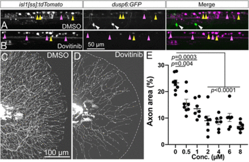
Dovitinib induces RB axon loss and reduction in Fgf signaling similar to SU5402 treatment. A,B, Lateral view of isl1[ss]:tdTomato; dups6:eGFP dorsal neurons in live 4 dpf larvae. Compared to DMSO-treated controls (A), short-term (7 h) dovitinib treatment (B) induced a substantial loss of Fgf signaling-dependent GFP expression. C,D, Lateral view of RB:GFP larval caudal tails at 5 pf following 48 h DMSO (C) or dovitinib (D) treatment. Dovitinib treatment led to major axon loss similar to genetic and pharmacological loss of Fgfr signaling. E, Quantification of dose-dependent effect of dovitinib on tail axon density. DMSO = 23.3 ± 1.1%, 0.5 μM = 15.7 ± 1.5%, 1 μM = 13.5 ± 1.3%, 2 μM = 9.0 ± 1.8%, 4 μM = 8.5 ± 1.0%, 6 μM = 10.2 ± 1.5%, 8 μM = 6.9 ± 0.6%, analyzed by one-way ANOVA with post hoc Tukey's test (F = 19.61, p < 0.0001). Yellow arrowheads = EGFP+ RB neurons, magenta arrowheads = EGFP− RB neurons, white arrowheads = EGFP+ motor neurons. |

