
Fig. 3
- ID
- ZDB-FIG-240524-55
- Publication
- Seliwjorstow et al., 2024 - Reversible Influence of Hemipiperazine Photochromism on the Early Development of Zebrafish Embryo
- Other Figures
- All Figure Page
- Back to All Figure Page
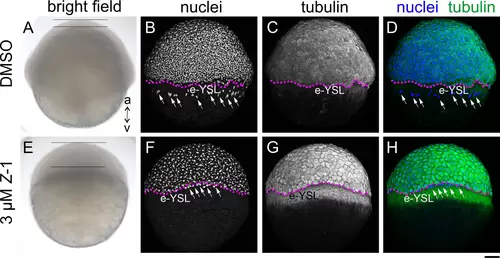
Treatment of Embryos with Z-1 Arrests Cell Division and Epiboly Movements. Phenotypic comparison between control (A-D, DMSO) and embryos treated with Z-1 (3 μM) (E-H) at 6 hpf, showcased through: (A and E) bright field view, (B and F; grayscale) DAPI nuclei staining, (C and G; grayscale) tubulin immunohisto staining, (D and H; nuclei in blue and tubulin in green) merged view of nuclei and tubulin; The animal pole (a) is oriented upwards, while the vegetal pole (v) is directed downwards. Notably, the Z-1-treated embryo (E) displays thickening of the blastoderm (span between two horizontal bars), a contrast to the control (A). Anomalous accumulation of tubulin is observed in the yolk syncytial layer (YSL) of Z-1-treated embryos, forming a belt-like structure vegetal to the blastoderm margin (stippled magenta line). The YSL nuclei (white arrows) were confined at the close vicinity of the blastoderm margin in the Z-1 treated embryo (F), while in the control they were found away from the margin and more in the vegetal region (B). Scale bar: 100 μm. |