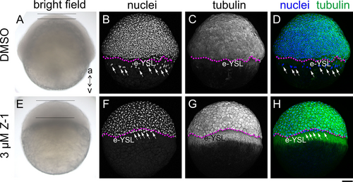
Fig. 3 Treatment of Embryos with Z-1 Arrests Cell Division and Epiboly Movements. Phenotypic comparison between control (A-D, DMSO) and embryos treated with Z-1 (3 μM) (E-H) at 6 hpf, showcased through: (A and E) bright field view, (B and F; grayscale) DAPI nuclei staining, (C and G; grayscale) tubulin immunohisto staining, (D and H; nuclei in blue and tubulin in green) merged view of nuclei and tubulin; The animal pole (a) is oriented upwards, while the vegetal pole (v) is directed downwards. Notably, the Z-1-treated embryo (E) displays thickening of the blastoderm (span between two horizontal bars), a contrast to the control (A). Anomalous accumulation of tubulin is observed in the yolk syncytial layer (YSL) of Z-1-treated embryos, forming a belt-like structure vegetal to the blastoderm margin (stippled magenta line). The YSL nuclei (white arrows) were confined at the close vicinity of the blastoderm margin in the Z-1 treated embryo (F), while in the control they were found away from the margin and more in the vegetal region (B). Scale bar: 100 μm.
Image
Figure Caption
Acknowledgments
This image is the copyrighted work of the attributed author or publisher, and
ZFIN has permission only to display this image to its users.
Additional permissions should be obtained from the applicable author or publisher of the image.
Full text @ Chembiochem

